Anatomy Of Musckes Sndctendons / Anatomy Muscle The Muscles The Tendons And Ligaments Are In White News Photo Getty Images / All together they help hold your upper arm in place in the shoulder.
Anatomy Of Musckes Sndctendons / Anatomy Muscle The Muscles The Tendons And Ligaments Are In White News Photo Getty Images / All together they help hold your upper arm in place in the shoulder.. See tendons muscles foot lower leg anatomy stock video clips. When the muscle contracts, the tendons are pulled, and the bone is moved. The muscles of the shoulder bridge the transitions from the torso into the head/neck area and into the upper extremities of the arms and hands. The wrist links the hand to the forearm. The legs include the upper leg, knee, lower leg, ankle, and.
The primary function of the shoulder girdle is to give strength and range of motion to the arm. The wrist joint is a complex joint which connects the forearm to the hand, allowing a wide range of movement. Tendons attach muscle to bone. Ebraheim's educational animated video describes the muscle anatomy of the hip and buttocks region with simple images; The peroneal muscles (peroneus longus and peroneus brevis), on the outside edge of the ankle and foot.
Shoulder Joint Anatomy Skeletal System Cartilages Ligaments Muscles Tendons from www.epainassist.com As these muscles contract and relax, they move skeletal bones to create movement of the body. All together they help hold your upper arm in place in the shoulder. The calf muscles (gastrocnemius and soleus), which are connected to the calcaneus via the achilles tendon. The knee joins the thigh bone (femur) to the shin bone (tibia). The smaller bone that runs alongside the tibia (fibula) and the. See tendons muscles foot lower leg anatomy stock video clips. Maybe you would like to learn more about one of these? The wrist joint is a complex joint which connects the forearm to the hand, allowing a wide range of movement.
Tendons also help to provide stability around the foot and ankle
When the muscle contracts, the tendons are pulled, and the bone is moved. The knee joins the thigh bone (femur) to the shin bone (tibia). Tendons also help to provide stability around the foot and ankle Maybe you would like to learn more about one of these? Anatomy of the hand and wrist: The wrist links the hand to the forearm. Each of them aids in a specific motion of your shoulder. One row connects with the ends of the bones in the forearm—the radius and ulna. Superficial muscles are the muscles closest to the skin surface and can usually be seen while a body is performing actions. The quadriceps are a collection of 4 muscles on the front of the thigh and are responsible for straightening the knee by bringing a bent knee to a straightened position. They act collectively to stabilise the arches of the foot, and individually to control movement of the digits. The primary function of the shoulder girdle is to give strength and range of motion to the arm. With the aid of numerous images, each chapter provides comprehensive descriptions of the anatomy, the normal mr appearance, pathological mr findings, and postoperative mri appearance.
Tendons vary in size and are somewhat elastic and attach bones to muscles. The wrist is a complex system of many small bones (known as the carpal bones) and ligaments. Schau dir angebote von muscle anatomy auf ebay an. We did not find results for: There are two main muscle groups around the knee:
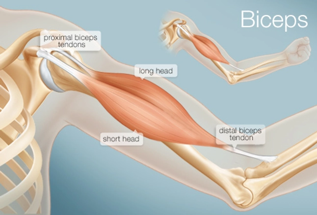
Human Muscle System Functions Diagram Facts Britannica from cdn.britannica.com The wrist links the hand to the forearm. Über 7 millionen englischsprachige bücher. This is lesson 1 on the anatomy of the forearm. There are a number of tendons located in the foot and ankle all responsible for different ankle, foot and toe movements. Maybe you would like to learn more about one of these? Although the majority of the muscle mass is located anteriorly to the humerus, it has no attachment to the bone itself. Ligaments and tendons are fibrous bands of connective tissue that attach to bone. Major muscles of the ankle.
Tendons also help to provide stability around the foot and ankle
Ligaments and tendons are fibrous bands of connective tissue that attach to bone. The muscles around the knee help to keep the knee stable, well aligned, and moving. Many in the neck help to stabilize or move the head. All the muscles are innervated either by the medial plantar nerve or the lateral plantar nerve, which are both branches of the tibial nerve. Wrist anatomy is the study of the bones, ligaments and other structures in the wrist. The muscles of the plantar aspect are described in four layers. The carpal bones are arranged in 2 interrelated rows. With the aid of numerous images, each chapter provides comprehensive descriptions of the anatomy, the normal mr appearance, pathological mr findings, and postoperative mri appearance. The smaller bone that runs alongside the tibia (fibula) and the. Tendons are thick bands of tissue that connect muscles to bone. The posterior upper leg muscles provide your knees with mobility (extension, flexion and rotation) and strength. Maybe you would like to learn more about one of these? On the other hand, the insertion is where a tendon attaches that muscle to the *more* movable bone.
Tendons also help to provide stability around the foot and ankle The quadriceps muscles provide strength and power with knee extension (straightening). Muscles of the neck (musculi cervicales) the muscles of the neck are muscles that cover the area of the neck hese muscles are mainly responsible for the movement of the head in all directions they consist of 3 main groups of muscles: The muscles you probably know the best are your. All together they help hold your upper arm in place in the shoulder.
The Biceps Human Anatomy Function Diagram Conditions More from img.webmd.com The muscles you probably know the best are your. Tendons also help to provide stability around the foot and ankle However, it is susceptible to injury, especially from repetitive strain. Maybe you would like to learn more about one of these? All the muscles are innervated either by the medial plantar nerve or the lateral plantar nerve, which are both branches of the tibial nerve. Superficial muscles are the muscles closest to the skin surface and can usually be seen while a body is performing actions. Ligaments connect two or more bones together and help stabilize joints. The gastrocnemius and soleus muscles taper and merge at the base of the calf muscle.
When a muscle contracts, the tendon pulls on the bone causing the joint to move.
Schau dir angebote von muscle anatomy auf ebay an. Many in the neck help to stabilize or move the head. Maybe you would like to learn more about one of these? The peroneal muscles (peroneus longus and peroneus brevis), on the outside edge of the ankle and foot. Menisci, cartilage, subchondral bone, patella, synovia, muscles and tendons, arteries, veins and bones. The calf muscles (gastrocnemius and soleus), which are connected to the calcaneus via the achilles tendon. We did not find results for: One row connects with the ends of the bones in the forearm—the radius and ulna. There are 10 intrinsic muscles located in the sole of the foot. The muscles of the plantar aspect are described in four layers. Anterior, lateral and posterior groups, based on their position in the neck.the musculature of the neck is further divided into more specific groups. The posterior upper leg muscles provide your knees with mobility (extension, flexion and rotation) and strength. The muscles you probably know the best are your.


0 Komentar